Home

Wasser Papua NeuGuinea Ventil stroke volume echo Mitternacht Geben Mission

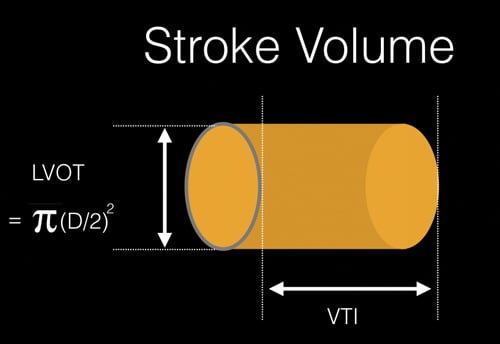
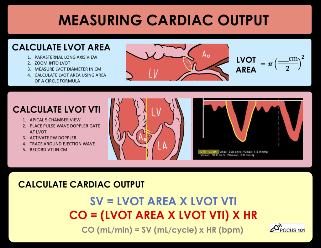
Measuring Cardiac Output with Echocardiography Made Easy - POCUS 101

Fig. 18. (A) Stroke volume by Doppler (LVOT). (B) Stroke volume by Doppler  (mitral inflow). (C)… | Diagnostic medical sonography, Cardiac sonography,  Echocardiogram
Fig. 18. (A) Stroke volume by Doppler (LVOT). (B) Stroke volume by Doppler (mitral inflow). (C)… | Diagnostic medical sonography, Cardiac sonography, Echocardiogram

3 Steps to Quantifying MR via Stroke Volume Method!

Normal Values of Cardiac Output and Stroke Volume According to Measurement  Technique, Age, Sex, and Ethnicity: Results of the World Alliance of  Societies of Echocardiography Study - Journal of the American Society
Normal Values of Cardiac Output and Stroke Volume According to Measurement Technique, Age, Sex, and Ethnicity: Results of the World Alliance of Societies of Echocardiography Study - Journal of the American Society

A, Normal LVOT VTI (VTI TSVI, 19.09 cm), indicating a normal stroke... |  Download Scientific Diagram
A, Normal LVOT VTI (VTI TSVI, 19.09 cm), indicating a normal stroke... | Download Scientific Diagram
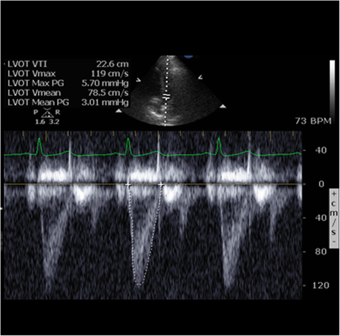
A novel method of calculating stroke volume using point-of-care  echocardiography | Cardiovascular Ultrasound | Full Text
A novel method of calculating stroke volume using point-of-care echocardiography | Cardiovascular Ultrasound | Full Text

Management of Shock: Bedside Assessment of Cardiac Output EMRA

Hemodynamics | Thoracic Key

Comparison of pulse pressure variation versus echocardiography-derived stroke  volume variation for prediction of fluid responsiveness in mechanically  ventilated anesthetized dogs - Veterinary Anaesthesia and Analgesia
Comparison of pulse pressure variation versus echocardiography-derived stroke volume variation for prediction of fluid responsiveness in mechanically ventilated anesthetized dogs - Veterinary Anaesthesia and Analgesia
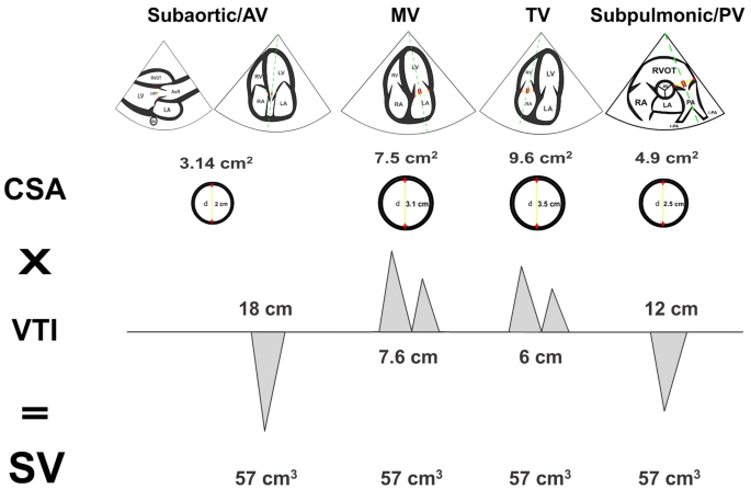
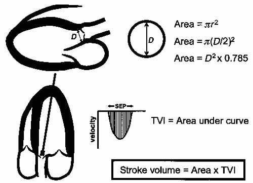
How To Calculate Stroke Volume In Echocardiography (The EASY Way)

Stroke Volume & Cardiac Output Measurement on POCUS Accurately ... | GrepMed

Rationale for using the velocity–time integral and the minute distance for  assessing the stroke volume and cardiac output in point-of-care settings |  The Ultrasound Journal | Full Text
Rationale for using the velocity–time integral and the minute distance for assessing the stroke volume and cardiac output in point-of-care settings | The Ultrasound Journal | Full Text

Stroke Volume, VTI (Velocity Time Integral) & Cardiac Output – ECG & ECHO

View Image
View Image

Accurate stroke volume (SV) estimation: SV = LVOT area × LVOT VTI. a... |  Download Scientific Diagram
Accurate stroke volume (SV) estimation: SV = LVOT area × LVOT VTI. a... | Download Scientific Diagram

Calculation of the stroke volume (SV) and cardiac output (CO) by... |  Download Scientific Diagram
Calculation of the stroke volume (SV) and cardiac output (CO) by... | Download Scientific Diagram

3 Steps to Quantifying MR via Stroke Volume Method!

Figure 1 from Automated quantification of mitral inflow and aortic outflow stroke  volumes by three-dimensional real-time volume color-flow Doppler  transthoracic echocardiography: comparison with pulsed-wave Doppler and  cardiac magnetic resonance ...
Figure 1 from Automated quantification of mitral inflow and aortic outflow stroke volumes by three-dimensional real-time volume color-flow Doppler transthoracic echocardiography: comparison with pulsed-wave Doppler and cardiac magnetic resonance ...

Point of Care Echo: Stroke Volume Determination - YouTube

Right Ventricular Stroke Volume from Doppler

Stroke Volume Determination by Echocardiography - CHEST

Estimation of Stroke Volume and Aortic Valve Area in Patients with Aortic  Stenosis: A Comparison of Echocardiography versus Cardiovascular Magnetic  Resonance - Journal of the American Society of Echocardiography
Estimation of Stroke Volume and Aortic Valve Area in Patients with Aortic Stenosis: A Comparison of Echocardiography versus Cardiovascular Magnetic Resonance - Journal of the American Society of Echocardiography
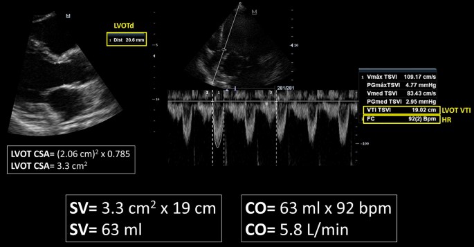
Management of Shock: Bedside Assessment of Cardiac Output EMRA

Rationale for using the velocity–time integral and the minute distance for  assessing the stroke volume and cardiac output in point-of-care settings |  The Ultrasound Journal | Full Text
Rationale for using the velocity–time integral and the minute distance for assessing the stroke volume and cardiac output in point-of-care settings | The Ultrasound Journal | Full Text
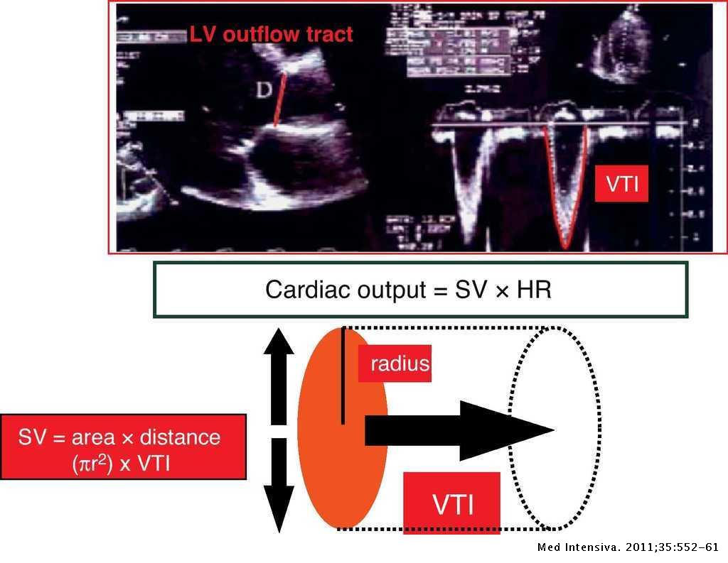
Estimating cardiac output. Utility in the clinical practice. Available  invasive and non-invasive monitoring | Medicina Intensiva
Estimating cardiac output. Utility in the clinical practice. Available invasive and non-invasive monitoring | Medicina Intensiva

Cardiac Output
Cardiac Output