Home

T. widerlich Neue Bedeutung cardiac echo measurements Einstellung Apfel Verstehen

THE AMERICAN SOCIETY OF ECHOCARDIOGRAPHY RECOMMENDATIONS FOR CARDIAC  CHAMBER QUANTIFICATION IN ADULTS: A QUICK REFERENCE GUIDE F
THE AMERICAN SOCIETY OF ECHOCARDIOGRAPHY RECOMMENDATIONS FOR CARDIAC CHAMBER QUANTIFICATION IN ADULTS: A QUICK REFERENCE GUIDE F

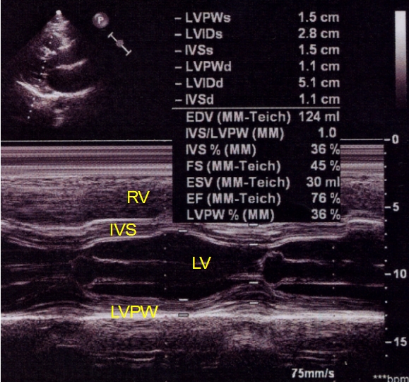
M-mode echocardiography measurement of the septum, posterior wall, LV... |  Download Scientific Diagram
M-mode echocardiography measurement of the septum, posterior wall, LV... | Download Scientific Diagram

Contrast-Enhanced Echocardiographic Measurement of Left Ventricular Wall  Thickness in Hypertrophic Cardiomyopathy: Comparison with Standard  Echocardiography and Cardiac Magnetic Resonance - Journal of the American  Society of Echocardiography
Contrast-Enhanced Echocardiographic Measurement of Left Ventricular Wall Thickness in Hypertrophic Cardiomyopathy: Comparison with Standard Echocardiography and Cardiac Magnetic Resonance - Journal of the American Society of Echocardiography

Small animal cardiac ultrasound measurements | IMV imaging

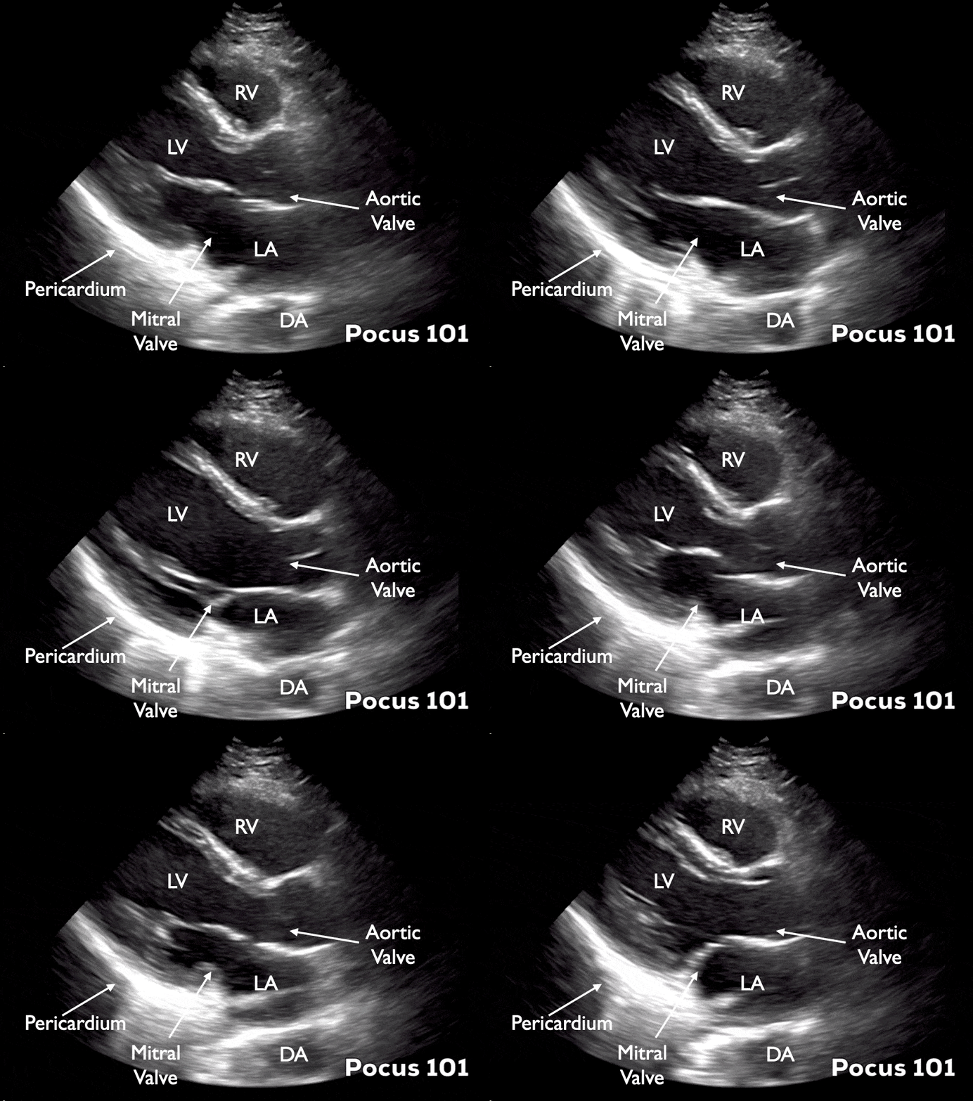
Measuring Cardiac Output with Echocardiography Made Easy - POCUS 101

Evaluation of automated measurement of left ventricular volume by novel  real-time 3-dimensional echocardiographic system: Validation with cardiac  magnetic resonance imaging and 2-dimensional echocardiography - Journal of  Cardiology
Evaluation of automated measurement of left ventricular volume by novel real-time 3-dimensional echocardiographic system: Validation with cardiac magnetic resonance imaging and 2-dimensional echocardiography - Journal of Cardiology

2D echo measurements needed for calculations of cardiac output... |  Download Scientific Diagram
2D echo measurements needed for calculations of cardiac output... | Download Scientific Diagram

Premature Closure Of Mitral Valve Picture - Echocardiography

Echocardiography — Cardiology Institute

M-mode (motion mode) echocardiography – ECG & ECHO

Measurement of cardiac output using transesophageal echocardiography.... |  Download Scientific Diagram
Measurement of cardiac output using transesophageal echocardiography.... | Download Scientific Diagram

M-Mode Echocardiographic Measurements – All About Cardiovascular System and  Disorders
M-Mode Echocardiographic Measurements – All About Cardiovascular System and Disorders

6 Pitfalls to Accurate LV Measurements #echo #echocardiography #CardioEd |  Medical ultrasound, Cardiac sonography, Echocardiogram
6 Pitfalls to Accurate LV Measurements #echo #echocardiography #CardioEd | Medical ultrasound, Cardiac sonography, Echocardiogram

Echocardiographic Analysis of Cardiac Function after Infarction in Mice:  Validation of Single-Plane Long-Axis View Measurements and the Bi-Plane  Simpson Method - Ultrasound in Medicine and Biology
Echocardiographic Analysis of Cardiac Function after Infarction in Mice: Validation of Single-Plane Long-Axis View Measurements and the Bi-Plane Simpson Method - Ultrasound in Medicine and Biology
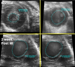
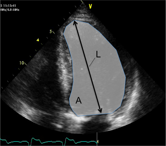
The Simpson's method for measurement of cardiac output.... | Download  Scientific Diagram
The Simpson's method for measurement of cardiac output.... | Download Scientific Diagram

High-frequency ultrasound measurement of heart function and fetoplacental  status using Vevo 770 | Cornell University College of Veterinary Medicine
High-frequency ultrasound measurement of heart function and fetoplacental status using Vevo 770 | Cornell University College of Veterinary Medicine
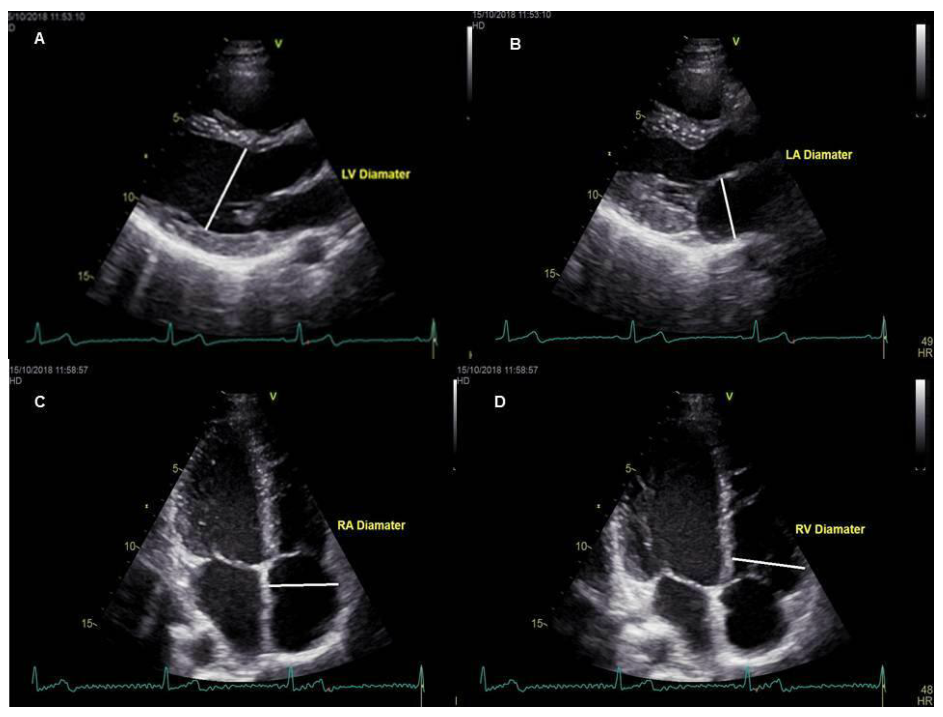
Medicina | Free Full-Text | Cardiac Magnetic Resonance Imaging and  Transthoracic Echocardiography: Investigation of Concordance between the  Two Methods for Measurement of the Cardiac Chamber | HTML
Medicina | Free Full-Text | Cardiac Magnetic Resonance Imaging and Transthoracic Echocardiography: Investigation of Concordance between the Two Methods for Measurement of the Cardiac Chamber | HTML
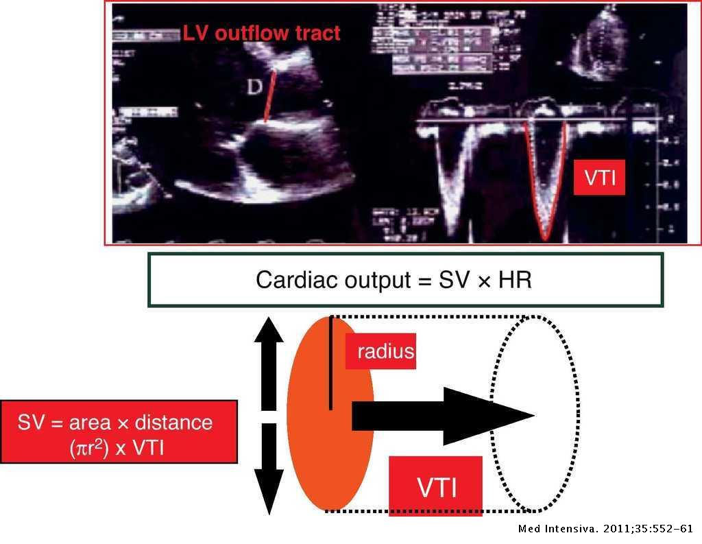
Cardiac Measurements from ECG Gated 2D Images | Download Table

Echocardiography in General Practice: 4 Views to Master | Clinician's Brief

Estimating cardiac output. Utility in the clinical practice. Available  invasive and non-invasive monitoring | Medicina Intensiva
Estimating cardiac output. Utility in the clinical practice. Available invasive and non-invasive monitoring | Medicina Intensiva

3D Quantitation
3D Quantitation

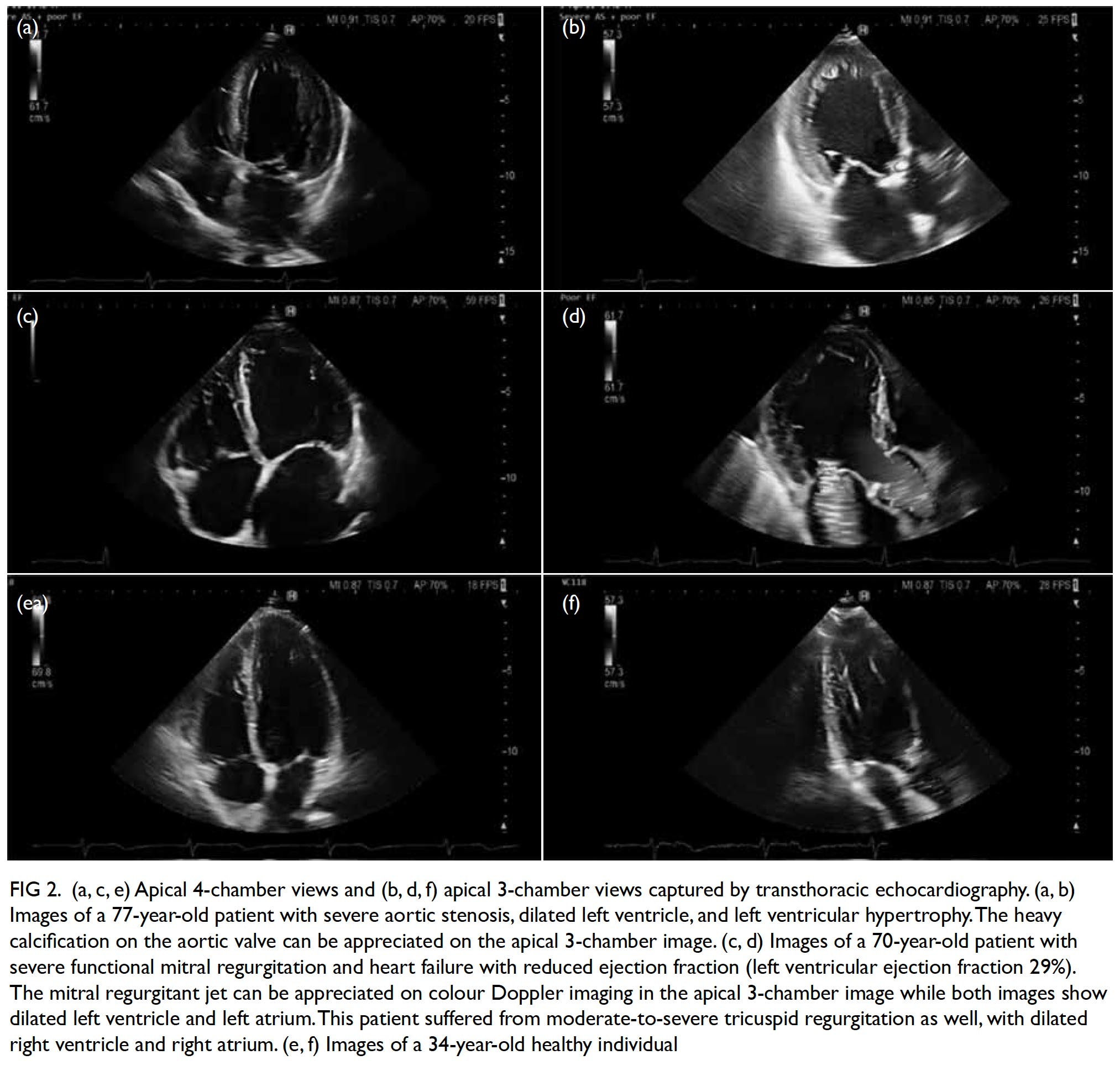
Echocardiography update for primary care physicians: a review | HKMJ

Fig. 18. (A) Stroke volume by Doppler (LVOT). (B) Stroke volume by Doppler  (mitral inflow). (C)… | Diagnostic medical sonography, Cardiac sonography,  Echocardiogram
Fig. 18. (A) Stroke volume by Doppler (LVOT). (B) Stroke volume by Doppler (mitral inflow). (C)… | Diagnostic medical sonography, Cardiac sonography, Echocardiogram
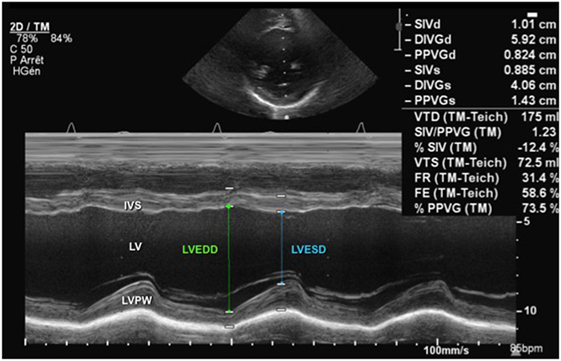
Frontiers | Echocardiographic Evaluation of Ventricular Function—For the  Neonatologist and Pediatric Intensivist | Pediatrics
Frontiers | Echocardiographic Evaluation of Ventricular Function—For the Neonatologist and Pediatric Intensivist | Pediatrics